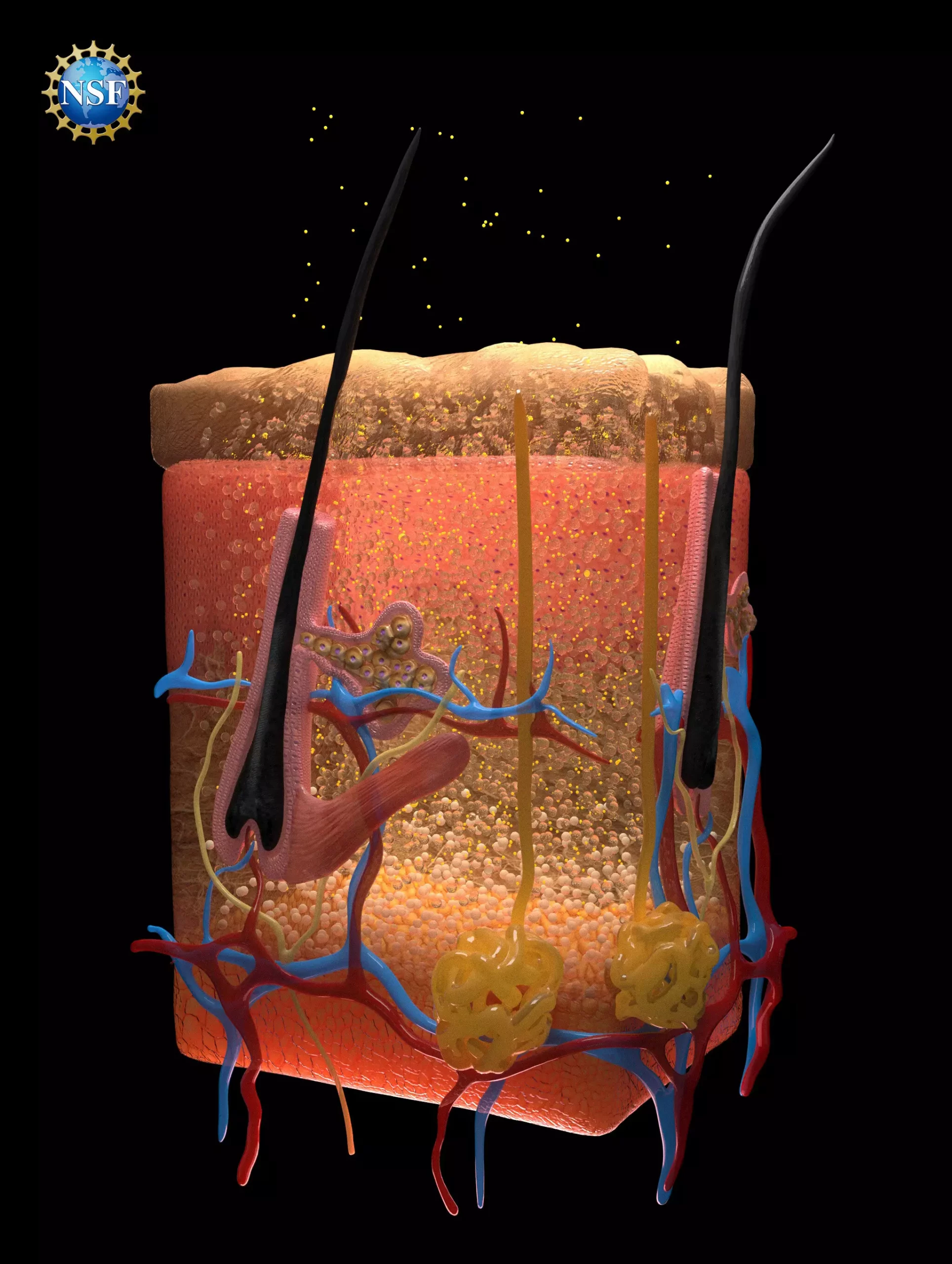
A transformative study emerging from Stanford University has unveiled a pioneering method of visualizing internal organs by rendering overlying tissues transparent to visible light. The innovative technique utilizes a food-safe dye that is applied topically, making it a reversible process. The implications of this research are vast, extending to various medical diagnostics, including the detection of injuries, monitoring digestive disorders, and identifying cancerous lesions. Published in the September 6, 2024, issue of *Science*, the study titled “Achieving optical transparency in live animals with absorbing molecules” showcases a significant advancement in medical imaging technology.
The researchers developed this technique by gaining insights into how light interacts with biological tissues, especially the complex ways it scatters and refracts as it moves through various materials. The body’s natural tissues consist of fats, fluids, proteins, and other biomolecules, each possessing distinct refractive indices—key properties that dictate how light bends and scatters. This scattering effect is what makes tissues appear opaque to our sight. To counteract this, the researchers sought to match the refractive indices of the dye with those of the tissues, enabling the light to pass through without disruption.
By leveraging fundamental principles from optics, such as light absorption and uniform penetration through materials, the team identified a specific dye, tartrazine, also known as FD & C Yellow 5, as particularly effective. When absorbed into biological tissues, it was found to align closely with the refractive indices of muscle fluids and proteins, effectively creating a transparent medium through which light could travel unimpeded.
Validation of this hypothesis began with the examination of thin chicken breast slices, where increasing concentrations of tartrazine resulted in a gradual transparency due to matched refractive indices. The success continued with live animal testing wherein researchers gently applied the dye solution to the scalp of mice, revealing intricate blood vessel networks in the brain, and later to the abdomen to observe intestinal contractions and cardiac activity. The ability to visualize such features at micron scales represents a significant leap in biomedical imaging, enabling enhanced microscopic observations without permanent alterations to biological tissues.
After the application was rinsed off, the tissues reverted to their standard opacity efficiently, and there were no adverse long-term effects observed in the test subjects, suggesting that the dye quickly cleared from the body, with any excess eliminated within 48 hours. The researchers speculate that by exploring injection methods for the dye, they may be able to achieve even deeper imaging capabilities, unveiling new insights into biological processes and medical conditions.
The project initially aimed to explore how microwave radiation interacts with biological tissues, a path that diverted into the realm of optics. The researchers drew upon classic optical theories, such as Kramers-Kronig relations and Lorentz oscillation, to reinforce their studies. Utilizing decades-old tools like ellipsometers—traditionally employed in semiconductor manufacturing, they discovered that these instruments are critical in predicting how various dyes impact the optical properties of tissues.
The team, which grew to include 21 students and collaborators, showcased a commendable effort in collaboration and innovation in medical imaging. The utilization of basic yet foundational instruments, such as the ellipsometer, illustrates how traditional scientific equipment can be repurposed to generate remarkable advancements in fields such as medicine.
The potential applications for this technology are extensive. It could simplify various procedures, such as drawing blood by making veins more visible or enhancing the effectiveness of laser-based treatments by improving light penetration. For oncological practices, it offers hope for earlier cancer detection, potentially changing the trajectory of diagnostics and treatment methods.
The transformative nature of this research highlights the importance of interdisciplinary approaches combining physics, biology, and engineering to forge innovations that can revolutionize medical practices. The method opens the door for future studies aimed at pairing dyes with biological materials, potentially birthing an entirely new field of study.
The development of techniques for rendering biological tissues transparent has significant promise for the future of medical diagnostics. This groundbreaking study encourages further exploration into optical sciences and its applications in the medical field, holding the potential to enhance diagnostic capabilities and improve patient outcomes dramatically. As researchers continue to refine these techniques, we may witness a new era in noninvasive imaging that profoundly impacts how medicine understands and treats complex conditions.

