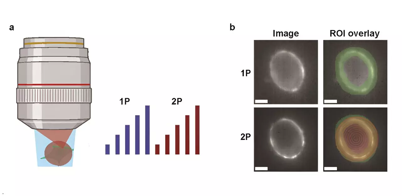
The brain, a complex network of neurons, orchestrates a multitude of functions through electrical signals. To decipher the intricacies of these signals, researchers have increasingly turned to genetically encoded voltage indicators (GEVIs). These indicators are revolutionizing how scientists visualize and understand neuronal activity. Notably, the discussion surrounding the efficacy of one-photon (1P) versus two-photon (2P) voltage imaging techniques has garnered considerable attention as researchers aim to unveil the principles governing neuronal communication.
A recent investigation conducted by a team at Harvard University offers a comprehensive comparative analysis between 1P and 2P voltage imaging methodologies. Published in the journal Neurophotonics, the study meticulously evaluates the optical and biophysical limitations inherent in each technique. Researchers concentrated on essential elements such as brightness and voltage sensitivity of various GEVIs utilized under both imaging modalities. A pivotal part of their investigation was also dedicated to analyzing fluorescence attenuation with depth in the mouse brain, a crucial consideration for in vivo studies.
To enrich their findings, the researchers constructed a predictive model that reflects the quantity of detectable cells based on various parameters, including imaging methods, reporter characteristics, and targeted signal-to-noise ratios (SNR). This quantitative approach provided a clearer framework for understanding the performance and practicality of these imaging techniques.
One significant revelation from the study is the stark disparity in power requirements between the two techniques. Specifically, the Harvard team discovered that 2P excitation necessitates about 10,000 times more illumination power per cell compared to 1P to achieve equivalent photon collection rates. This distinction raises pressing concerns regarding potential tissue damage and the noise generated during imaging. For instance, deploying the JEDI-2P indicator in the mouse cortex at an SNR of 10 restricts the simultaneous measurement capability to approximately 12 neurons at depths exceeding 300 micrometers, given the constraints of laser power and frequency.
These findings underscore a critical hurdle: the current limitations of 2P voltage imaging in vivo reveal that achieving sufficient SNR for hundreds of neurons at considerable depths remains a formidable challenge. The study emphasizes that enhancements in the capabilities of 2P GEVIs, or the invention of novel imaging strategies altogether, are essential to propel advancements in this research area.
As scientists continue to navigate these complexities, the insights gleaned from the Harvard study serve as a guiding beacon for future endeavors in the field of neural imaging. The interplay between 1P and 2P techniques highlights the necessity for ongoing innovation and advancement in imaging technologies, as researchers strive to deepen our understanding of the electric symphony that governs neuronal interactions. The future of neuroscience hinges on overcoming these imaging obstacles, ultimately paving the way for breakthroughs in understanding neural circuits and their functionalities.

