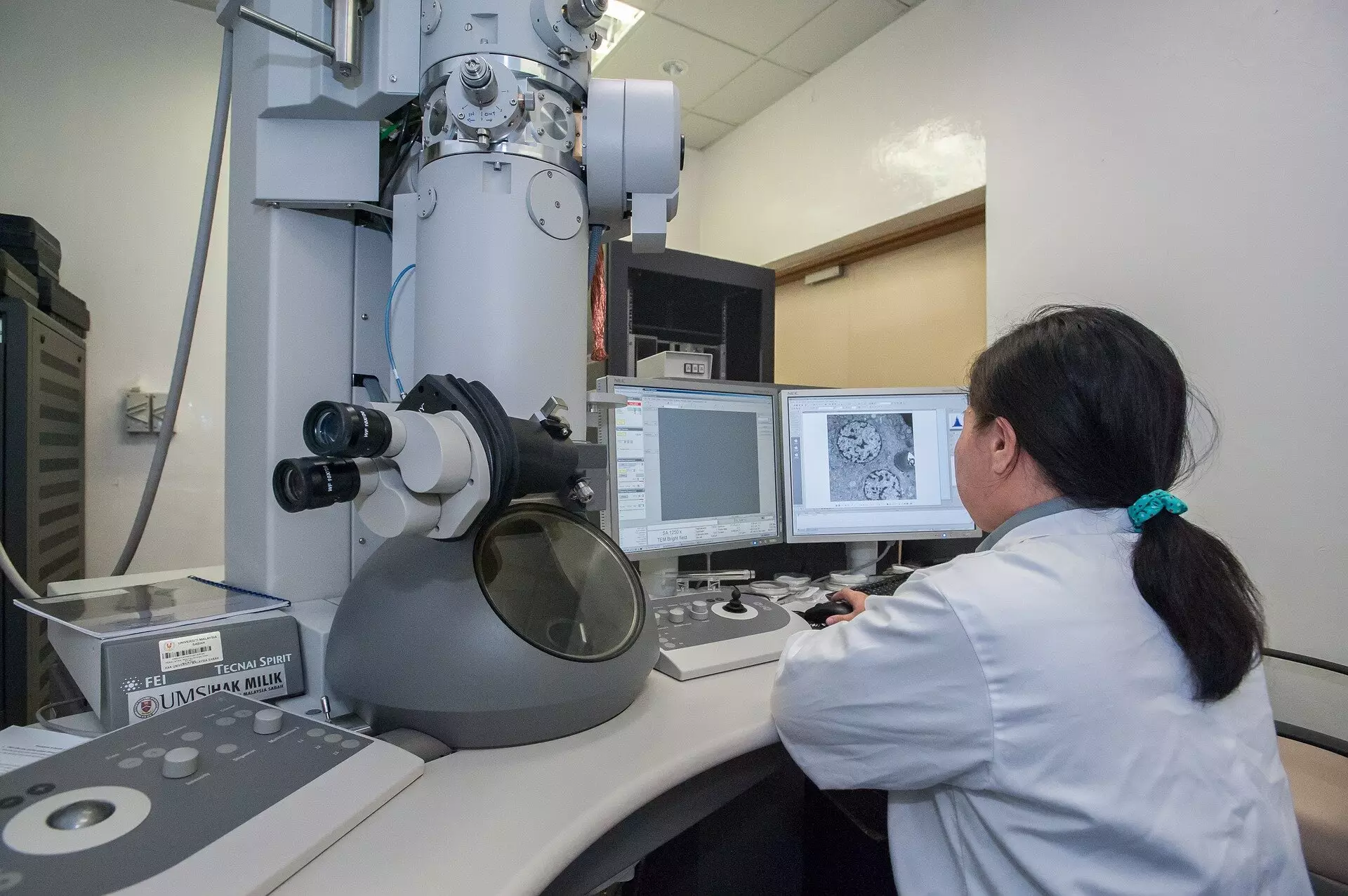
In the rapidly evolving field of microscopy, technological advancements continually reshape the landscape of scientific discovery. The latest breakthrough emanating from an international collaboration, spearheaded by researchers at Trinity College Dublin, introduces a pioneering imaging method that promises to redefine how we capture images at the microscopic level. This innovation not only lowers the time needed for imaging but also significantly reduces the detrimental radiation exposure to delicate samples, particularly critical in biomedical applications.
Traditional scanning transmission electron microscopy (STEM) relies on a methodical sequential approach, where an intense beam of electrons traverses the sample point by point. While this conventional method has served the scientific community well, it introduces an inherent risk: the potential for damaging sensitive samples due to prolonged exposure to electron radiation. The straightforward nature of fixed dwell times has led to high radiation doses, which, rather than enhancing image quality, can compromise the integrity of the biological tissues or materials being examined. For researchers, this poses a serious challenge, especially when the objective is to glean accurate information from fragile samples.
Event-based Detection: A Paradigm Shift in Imaging
The cutting-edge imaging method proposed by the Trinity team represents a seismic shift in the theoretical approach to microscopy. By abandoning the conventional fixed-exposure model, the researchers have embraced an event-based detection system. Instead of waiting for a predetermined dwell time for each pixel, this innovative system measures the time taken to detect a specified number of scattering events. This mathematical recalibration supports the assertion that the first electron detected at a given position carries a wealth of information, whereas subsequent detections yield diminishing returns.
This recalibration in logic and methodology could herald a new era for carcinogenic materials and delicate subjects within various fields—particularly materials science and medicine, where precision is paramount. Researchers have long grappled with the challenge of balancing image quality and sample preservation. The new approach effectively brings the focus back to maximizing imaging efficiency while minimizing destructive radiation exposure.
Tempo STEM: The Future of Safe, High-Quality Imaging
To actualize this theoretical foundation, the research team has developed and patented an ingenious technology dubbed Tempo STEM, in collaboration with IDES Ltd. This advancement incorporates an advanced beam blanker that allows for real-time control of the electron beam—shutting it off moments after sufficient data has been acquired. The ramifications for microscopy practices are profound; microscopists now possess the capability to dynamically adjust the beam operations, providing exquisite control over imaging conditions.
Dr. Lewys Jones, the lead investigator of this groundbreaking work, articulated the monumental impact of this combined technology. He highlighted how the capability to shutter the beam in nanoseconds in response to real-time events represents an unprecedented leap in microscope functionality. This newfound ability reduces the overall radiation dose without compromising the quality of the resulting images, a significant achievement considering the complexities of capturing high-resolution images of minute biological specimens. The importance of this advancement cannot be overstated; avoiding unnecessary damage to samples opens new avenues for research and clinical applications, pushing the boundaries of what can be achieved using electron microscopy.
Broader Impact and Future Perspectives
As the doors of possibility swing open due to this innovative imaging technique, the implications stretch far beyond the realms of traditional microscopy. Improved imaging of biological tissues can significantly influence how disease mechanisms are understood, paving the way for potentially more accurate diagnostics and targeted therapeutic approaches. The advancement also extends to materials science, where understanding the microscopic behavior of materials can lead to the development of better products—ranging from stronger composites to safer pharmaceuticals.
Dr. Jon Peters, the first author of the study, underscored the significance of reducing radiation levels in electron optics. He pointed out that while electrons are often viewed as benign, their high-speed impacts can lead to substantial damage within biological samples. This capability to minimize destructive doses while maintaining image fidelity is instrumental for scientists aiming to gather precise data without the trade-off of sample viability.
The groundwork laid by the Trinity College Dublin team has the potential to ripple through multiple scientific disciplines, enhancing our understanding of the natural world. With innovative systems like Tempo STEM leading the way, the future of microscopy appears not only brighter but also much safer.

