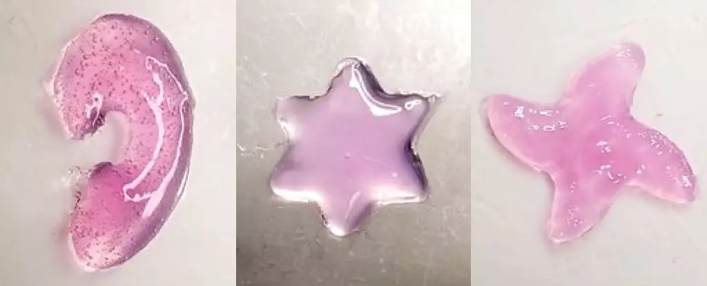
Recent strides in biomedical engineering are pushing the boundaries of traditional medicine, unlocking novel treatment avenues for diseases and injuries. The latest breakthrough comes from a team of scientists at the California Institute of Technology (Caltech), who have developed a method to 3D print materials directly within the body using ultrasound technology. This innovative approach, dubbed deep tissue in vivo sound printing (DISP), marks a significant leap in how we conceive of surgical procedures and therapeutic interventions, particularly in fields like oncology and regenerative medicine.
Not only does this technique have the potential to administer localized cancer treatments, but it could also open doors to repairing damaged tissues more efficiently and accurately than previous methods allow. The essence of DISP lies in using a specialized bioink, a concoction that varies based on therapeutic objectives, but its foundational components consist of polymer chains and crosslinking agents. These agents are crucial because they enable the formation of a hydrogel—a soft, gel-like material that can serve multiple purposes within the body.
The Science Behind DISP: Mechanisms of Action
To prevent the hydrogel from forming immediately upon injection—an imperative for effective implementation—the researchers encapsulate the crosslinking agents within lipid-based particles known as liposomes. These liposomes are designed to release their contents when exposed to specific temperatures, in this case, slightly above normal body temperature. Once the ultrasound beam is directed at the target area, the localized heating causes the liposomes to rupture, releasing the crosslinking agents and enabling the formation of the hydrogel in real-time.
What differentiates DISP from previous methodologies is its ability to penetrate deeper tissues. While infrared light has been utilized in the past for 3D printing, its effectiveness is limited to the surface layers of the skin. In contrast, ultrasound can reach deeper into muscles and organs, providing a significant advantage that expands the scope of potential applications. The control that researchers achieve by modulating the ultrasound beam facilitates the printing of intricate and versatile structures, which bodes well for treating a myriad of health conditions.
The Multifaceted Applications of Hydrogel Printing
The practical uses of DISP extend far beyond mere cosmetic enhancements. During preliminary tests with hydrogel variations, the team demonstrated the capacity to replace or regenerate damaged tissues and administer vital pharmaceuticals. Moreover, the technology allows for the monitoring of bodily functions, such as electrical signals during electrocardiography tests.
One exciting aspect of DISP is its capability to incorporate gas vesicles as imaging contrast agents, offering real-time monitoring of the printing process. By observing changes in contrast due to the chemical reactions instigated by polymer crosslinking, researchers can confirm the successful deployment of the hydrogel. This feature is especially relevant in a clinical setting where precision is paramount.
Additionally, trials in rabbits showcased the ability to print tissue constructs at depths of up to 4 centimeters beneath the skin, paving the way for expedited healing processes. Most impressively, the engineered hydrogel enabled targeted drug delivery, as illustrated in studies involving bladder cancer. When loaded with doxorubicin—a common chemotherapeutic agent—DISP facilitated prolonged and controlled drug release, resulting in increased efficacy and cancer cell death compared to conventional methods.
The Future of Biomedical Engineering: Bridging the Gap to Human Applications
While the success of DISP in animal models is promising, the transition to human clinical trials remains a monumental challenge. Researchers must address several crucial factors, including ethical concerns, regulatory approvals, and ensuring biocompatibility. The Caltech team remains optimistic as they plan to move to larger animal models before potentially embarking on human evaluations.
The exploration of deep tissue in vivo sound printing unlocks an exciting chapter in the realm of medical technology. The ability to conduct intricate, localized treatments directly within the body could redefine how we approach injury recovery and disease management, promising a future where healthcare is not just reactive but profoundly proactive. As researchers like Wei Gao continue to pioneer such innovations, we may be on the brink of a seismic shift in patient care and therapeutic strategies.

